For this post I have a loupe with two different powers. This is a low cost model, but has decent glass optics. A single ended loupe of 10X to 20X would be the Bausch and Lomb Coddington (2 element) or Hastings Triplet (3 elements).
In these photos, the loupe unit is tacked onto the phone with "blue tac" (Loctite Fun-Tak).

In the first photo, the lens is swung out, so it is lined up with the camera lens.
In the second photo the loupe lens is swug away, exposing the camera lens, so to be able to shoot normally.

This is the stuff: Loctite Fun-Tak.
Some shots with various loupes
 |
| Without loupe |
Bouganvilla: a small shift in focus plane makes a big difference.

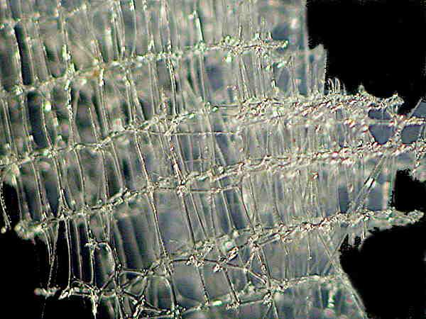
 |
| Very small flower |
 |
| A spittle bug in California |
 |
| On a lemon leaf in CA |
Tricks/Thoughts:
- when shooting, shoot in bursts, and move the camera in and out a little, to change the plane of focus.
- It is best to shoot in bright sunlight. I still haven't figured out how to use the onboard flash with this method. Must use either a diffuser or some kind of minature slave flash.
VideoFlex

I found a classic VideoFlex. As originally built, the VideoFlex gooseneck microscope and macro camera was a low resolution solution to display on a TV. This the one I got on Ebay for about 30.00. I found it invaluable in the classroom for displaying demos. My class watched a
Conus striatus attack and kill a blenny in an 8" culture dish, a definite highlight.
But the most interesting trick was the use of the VideoFlex as an Aquarium microscope. I had made some miniature aquaria of various sizes, including my favorite 2"wide X 2" long X 8" high, of 1/8" plate glass. This was a perfect size to empty a partly full 1 gallon ziploc bag of water (sea water, of course) and whatever specimens I had collected. These small aquaria were made by my predecessor instructors at Northern Marianas College to hold fish for photography. I found them useful for another reason.
On the sand in shallow water on Saipan are patches of a brown surface film. Curious, I used the ziploc bag like a scraper, and carefully scraped off a good amount of the surface film, as thinly as possible so as to avoid collecting deeper sand; the bag was filled the rest of the way with seawater. Usually, this about 2-1/2 or 3" deep worth of sand in the aquarium, which also held most of the water that I collected with it.

It took a little while for the organisms that had made up the surface layer---the epifauna, I suppose---to start reorganizing themselves in some manner. When the VideoFlex camera was placed flat on the surface of the aquarium, exactly along the surface line of the sand, one could watch this reorganization happen, as worms started to burrow, and, more interestingly, perhaps, dinoflagellates actually were resolved as they crawled up the glass into the water column above the sand. Fortuitously---depending on one's point of view---the dinoflagellates I observed were
Prorocentrum, which is toxic. The resolution wasn't great. It occurs to me that other cameras may be even more fit for this purpose; however, the VideoFlex's lens is the exact right focal length to focus on the inside surface of the glass, when the face of the lens if flush with the outside surface of the 1/8" glass. Awesome form meiofauna/infauna studies at the macro level.
Along with the 35.00 used (in good condition) VideoFlex came two power supplies, and an adaptor for a certain size of microscope eyepiece often used on stereomicroscopes.
I have attached the adaptor to the phone, making it much easier to line up for shots. I have not tried this adaptor on my compound scope, whose eyepiece is smaller in diameter.
This is a shot of a "worm" Fe found in the water residue on the back of the kitchen sink, using the iPhone camera through a stereomicroscope, using the collar tacked onto the case of the phone:
All in all, the cell phone camera has started to get interesting as a serious tool.
UPDATE: This system has its limits, esp. with regard to distortion.
These photos show spherical aberation. Should have paid better attention in Optics Class: should be able to construct a multiple element system with a few elements from Edmund Optical, or perhaps find an element to attach to one of these.
Maybe spacing of lens from camera?
 |
| Lichen. I assume this is a reproductive structure. |
|
Anyway, these shots still are interesting.
 |
| Elysium, a very small flower. | | |
|